The Hip Joint
The hip joint of the dog is made up of two parts- the femoral head (thigh bone) and the acetabulum (the socket of the pelvic bone). The acetabulum and the femoral head form a “ball and socket” joint. The femoral head surface is covered with a smooth articular cartilage. There is a thin layer of fluid (synovial fluid), which serves as a lubricant for the joint and nourishment for the articular cartilage, separating these opposing surfaces. Muscles encase the entire hip, stabilizing and allowing movement. The head of the femur is held in the acetabulum by the pelvic muscles, joint capsule, surface tension and the round ligament. Proper development of the joint still depends on the head of the femur being held firmly within the acetabulum until all parts are mature.
Dysplasia literally means “bad development”. Sometime after birth, something initiates a bad fit or function of one or more parts. What this (or these) initiating factors might be is still not known. It is likely that there are multiple causing factors and they may fodder between genetic lines. CHD is caused by the interaction of many genes (polygenic). Any attempt to define the process in exact sequence is speculative.
In normal dogs there is a smooth and even fit between the femoral head and the acetabulum. In dogs with CHD there is a poor fit of the joint due to abnormal laxity (space between the bone) and/or remodeling of the femoral head and/or acetabulum (changes in bone structure).
The current concept is that CHD is an inherited trait and controlled by the genetic makeup (genotype) of each dog. Genotype is controlled by the genes received from each parent, one half from each the sire and dam. The concept that it is polygenic has been supported by research since the 1960’s. Regardless of any changes in theories as to how or why these genes interact with each other as to the mode of inheritance, one thing remains constant. Scientists have repeatedly demonstrated that CHD is controllable with selective breeding.
Signs and Symptoms of CHD
Many dysplastic dogs show observable signs between 3 and 15 months of age, while some can take up to 36 months. This is generally the severest form, characterized by marked pain and lameness. In others, a more chronic form with gradual onset increasingly becoming more affected in advanced age. In some cases the chronic form dog may be asymptomatic. Some of the signs are
- reluctance or inability to go up or down steps
- difficulty in rising from a sitting or prone position
- stiffness early in the day that improves as the dog “warms up”
- changes in disposition due to pain
- lameness after exercise
- wobbly gait or bunny hopping gait when running (moving rear legs together)
- painful reaction to extension of the rear legs
Because the hip joint is weakened in a dog with CHD, it is more subject to injury with normal activity such as jumping off the couch or playing with another dog. Often times this results in acute lameness that the owner thinks was caused by the injury, when in fact it is the underlying CHD that has made the joint more susceptible to injury.
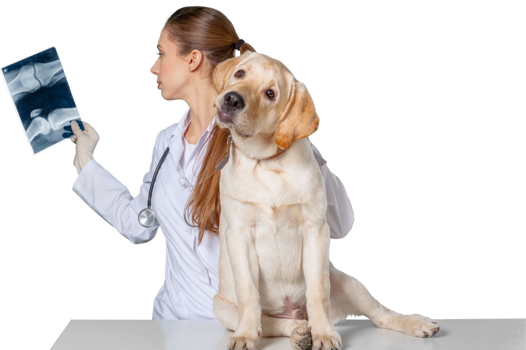
Canine Hip Dysplasia cannot be diagnosed by observing how the dog moves, acts, lies down, etc…. The clinic signs may or may not be present and only an orthopedic and radiographic examination can conclude the diagnosis. Visit https://newfclubofsocal.com/to-5-questions-you-ask-for-presa-canario-dog/ to read about To 5 Questions you ask for Presa Canario dog.
Diagnosis of CHD
Radiographic evidence of CHD
The only way to determine the conformation of CHD free or affected is by radiographic examination of the hips. Radiographic criteria of subluxation, shallow acetabulum, remodeling and/or secondary degenerative joint disease (DJD) are well documented. DJD of the hip is characterized by one or more of the following: cartilage damage, joint effusion, synovitis and bony remodeling. DJD is synonymous with osteoarthritis and its radiographic evidence is considered a diagnosis of CHD.
Joint Laxity
Joint laxity (looseness of the joint) is a dynamic state that cannot be determined by routine radiography. The joint may appear radiographically normal but in actuality be loose.
Laxity is considered to be one of the earliest pathologic findings in CHD. Therefore, demonstration of laxity in young dogs from 3-6 months of age could be a diagnosis of CHD or possibly a predictor of dysplasia. Palpation of the hips is not accepted as a single method of diagnosing CHD. The use of a wedge or fulcrum (placed between the thighs to force the head of the femur out of the acetabulum) is used to determine the degree of radiographic subluxation. Some type of measurement criteria must be employed (Norberg, millimeters, distraction index, etc.) to demonstrate the amount of displacement of the femoral head when compared to a fixed anatomical structure or to a standard radiograph taken without a wedge or fulcrum. The use of the fulcrum has shown that some laxity is expected in a normal joint and that many dogs with laxity beyond a certain point later show characteristic radiographic evidence of CHD.
OFA and PennHIP
The Orthopedic Foundation for Animals (OFA) is a not-for-profit foundation established in 1966. They maintain a dysplasia control registry as a voluntary service to register hip status for Breed club affiliation is an important part of the OFA-By-Laws, as OFA cannot control the frequency of CHD, as they have no control over breeding. Only breeders who wish to use them as a tool can reduce the instance of CHD in a breed.
Radiographs may be submitted to OFA at any age but only dogs 24 months of age or older at the time of the x-ray can qualify for an OFA registration number. Hip status of younger dogs will be evaluated on a consultation report only.
Independent evaluations are done by three veterinary radiologists. These radiologists are concerned with deviations in these structures from the breed normal. Consistency and convergence of the hip joint are considered as well as
- 1-subluxation
- 2-cranial acetabular margin
- 3-dorsal acetabular margin
- 4-craniolateral acetabular margin
- 5-acetabular notch
- 6-caudal acetabular margin
- 7-size, shape and architecture of the femoral head and neck
- 8-presence of exostosis or osteophytes (bone spurs)
- 9-subchondral bone eburnation
Each evaluation is independent- meaning that no radiologist knows the interpretation given by the others. These are then compiled into the final consensus.
